
Radiology & Interventional Neuroradiology
Radiology is a medical specialty that employs the use of imaging to both diagnose and treat disease visualised within the human body. Radiologists use an array of imaging technologies such as X-ray radiography, ultrasound, computed tomography (CT), nuclear medicine, positron emission tomography (PET) and magnetic resonance imaging (MRI) to diagnose or treat diseases. Interventional radiology is the performance of (usuallyminimally invasive) medical procedures with the guidance of imaging technologies. The acquisition of medical imaging is usually carried out by the radiographer or radiologic technologist. The radiologist then interprets or “reads” the images and produces a report of their findings and impression or diagnosis. This report is then transmitted to the ordering physician, either routinely or emergently.
Computed Tomography (CT)
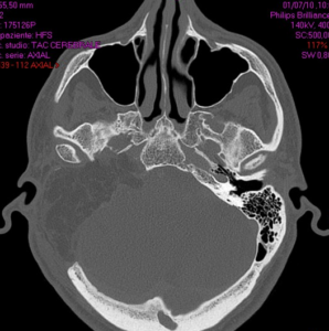
CT imaging uses X-rays in conjunction with computing algorithms to image the body. In CT, an X-ray tube opposite an X-ray detector (or detectors) in a ring-shaped apparatus rotate around a patient, producing a computer-generated cross-sectional image (tomogram). CT is acquired in the axial plane, with coronal and sagittal images produced by computer reconstruction. Spiral multidetector CT uses eight, 16, 64 or more detectors during continuous motion of the patient through the radiation beam to obtain much finer detail images in a shorter exam time. With rapid administration of intravenous contrast during the CT scan, these fine detail images can be reconstructed into three-dimensional (3D) images of carotid, cerebral, coronary or other arteries.
Magnetic resonance imaging (MRI)

MRI uses strong magnetic fields to align atomic nuclei (usually hydrogen protons) within body tissues, then uses a radio signal to disturb the axis of rotation of these nuclei and observes the radio frequency signal generated as the nuclei return to their baseline states. The radio signals are collected by small antennae, called coils, placed near the area of interest. MRI scans give the best soft tissue contrast of all the imaging modalities. With advances in scanning speed and spatial resolution, and improvements in computer 3D algorithms and hardware, MRI has become an important tool in musculoskeletal radiology and neuroradiology.
Interventional Radiology & Neuroradiology
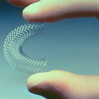
Interventional radiology (IR or sometimes VIR for vascular and interventional radiology) is a subspecialty of radiology in which minimally invasive procedures are performed using image guidance. Some of these procedures are done for purely diagnostic purposes (e.g., angiogram), while others are done for treatment purposes (e.g., angioplasty). The basic concept behind interventional radiology is to diagnose or treat pathologies, with the most minimally invasive technique possible. Interventions in the head and neck can be performed via percutaneous, endovascular, or a combination of these approaches. Procedures that predominantly require percutaneous access include biopsies and aspirations, sclerotherapy and newer techniques like radio-frequency ablation and cryoablation. On the other hand, a transarterial (or endovascular) approach forms the mainstay of treatment for head and neck bleeding as well as for transarterial chemotherapy for head and neck neoplasms. A combination of percutaneous and transarterial approaches may be needed in the embolization of high-flow craniofacial vascular malformations (VMs) and hypervascular tumors.
